Researchers and clinicians have been studying the biological behaviour of cells and tissues to understand, diagnose and treat diseases and health conditions. It is quite recently that researchers have started to observe how the physical and mechanical properties such as elasticity, stiffness and strength of cells and tissues affect body processes in health and disease.
Stiffness of a cell, an indicator of how much it deforms under applied force, is seen to change in different diseases and conditions. For example, the stiffness of red blood cells may increase due to ageing or in diseases such as malaria and sickle cell disease and the stiffness of cancer cells is observed to reduce compared to the normal cells of the same kind. The ability to measure the stiffness of cells can potentially help in the early diagnosis of certain diseases, evaluating the current state of the disease and prognosis, and finding drug candidates for disease treatment.
Savita Kumari, Ninad Mehendale and Prof Debjani Paul of the Indian Institute of Technology Bombay (IIT Bombay) and Prof Dhrubaditya Mitra of the Nordic Institute for Theoretical Physics, Sweden have developed a microfluidic device that can measure the stiffness of thousands of red blood cells (RBCs) in human blood in a few seconds. The research was published in the journal Cell Reports Physical Science.
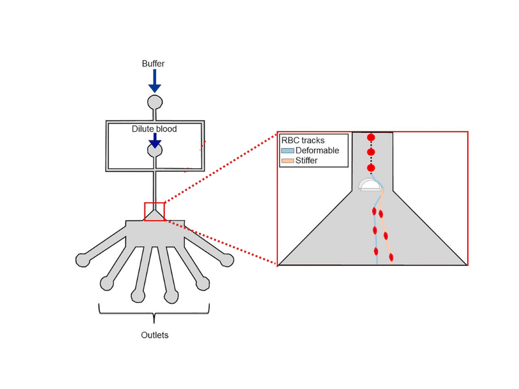
The IIT Bombay device is compact and portable. It is easy to use at point-of-care to monitor RBC stiffness in blood samples of patients with sickle cell disease or malaria. RBCs in stored blood bags can also sometimes become stiff, making the blood unusable for transfusion. The device can also be used to conduct a quick and easy assessment of the stored blood before transfusion to ensure that a bad bag of blood is not transfused. The device uses a tiny microfluidic chip and a portable optical microscope. An analysis software scans the video captured by the microscope to give a distribution of the stiffness of RBCs in the blood sample.
Conventional methods use various approaches to measure the stiffness or deformability of cells. However, most of them rely on measuring the deformability of a single cell at a time. These methods are expensive and time-consuming, and some methods like optical tweezers, optical stretchers and atomic force microscopy need apparatus as big as a wardrobe. Other microfluidics-based methods rely on observing how the cell changes its shape. Hence, these methods require a high-speed camera capable of capturing 3000 frames per second, at least 100 times faster than a consumer digital camera.
“Our device utilises a unique device design to probe the specific behaviour of soft deformable structures, in this case cells, flowing in a liquid medium. When the width of the channel through which cells are flowing changes, the forces acting on the cells change, and depending on the stiffness of the cells their paths through this device change,” explains Prof Debjani Paul, the lead researcher of the study.
The IIT Bombay device has a channel a few tens of micrometres wide (about as thick as a strand of human hair) through which the RBCs can flow. It opens into a funnel. A semi-cylindrical column is present as an obstruction at the junction of the channel and the funnel. When the steadily and rapidly flowing RBCs encounter the obstacle, they get deflected as they enter the funnel. The stiffer they are, the wider they are deflected. The stiffness can be computed based on the deflection of the RBCs.
A short video of the RBCs passing through the IIT Bombay device, played at half the speed.
Credit : Savita Kumari and Debjani Paul
As crucial as it is to validate a concept theoretically, making an affordable and simple-to-use working device needs careful engineering.
“It was challenging to fabricate a funnel whose width is ten times its height. It would collapse during fabrication if we were not very careful,” says Prof Paul.
The channel is just 5 micrometres tall to ensure that the RBC stays flat as it approaches the semi-cylindrical column. The channel width is 40 micrometres and the cone of the funnel opens wider, whereas the height of the conical portion is maintained at 5 micrometres.
The measure of stiffness is a quantity called Young’s modulus. The video recorded from the microfluidic device-microscope-camera setup records trajectories of the RBCs from which we can get the deflection angles. The deflection angles given by the device need to be mapped to the corresponding Young’s modulus value. In a one-time calibration exercise, the team measured deflection angles for healthy RBCs and mapped the deflection angles to Young’s moduli of the RBCs from the same sample measured using an atomic force microscope (AFM). Tanusri Roy, led by Prof Shamik Sen (both IIT Bombay), performed the AFM measurements.
Ideally, one would send a single RBC through the IIT Bombay device, measure Young’s modulus of the same RBC using an AFM, and repeat this hundreds of times to calibrate the device. Given the nature of the experiments involved, the single RBC method is practically impossible. The team used a data-based approach to calibrate the device. Prof Dhrubaditya Mitra led the algorithm development that mapped the value of the deflection angle of the RBCs to its Young's modulus.
“The algorithm gave us a calibration curve that can now be used to directly relate the deflection angle of any unknown RBC to its stiffness, without needing any other additional measurement such as atomic force microscopy,” says Prof Mitra.
The researchers validated the microfluidic device using sets of artificially stiffened RBCs.
Though the device can currently be used to measure the stiffness of RBCs only, it can be made about 100 times smaller to measure the stiffness of nano-sized objects, such as extracellular vesicles. Extracellular vesicles are just visible as bright point sources under high-resolution microscopes. Methods that rely on changing shape to measure stiffness cannot be used to measure their stiffness.
“As our method relies on measuring the trajectory of the elastic object and not its shape, it is more easily generalisable to such nanoscale objects,” comment the authors.
The team plans to develop the device to measure the stiffness of other types of cells in the body and handle cells of different shapes and sizes.