

Researchers at the Indian Institute of Science (IISc) here have designed a novel device that can help biologists to stretch cells and study them one at a time.
“The best part about this device is that scientists can see and analyze cells in real time under the microscope”, says Mr. Sreenath Balakrishnan from the Center for Biosystems Science and Engineering (BSSE) at IISc, one of the researchers involved in the study. A culmination of efforts from researchers at the Department of Mechanical Engineering (ME), BSSE, and the Department of Microbiology and Cell Biology at IISc, this study led by Prof. Ananthasuresh, a Professor in the BSSE and ME departments at IISc, was published in the Journal of Micro-Bio Robotics.
Human body, made up of about 3 × 10¹³ (or 30 trillion) cells, picks up different cues and responds to various ‘signals’ in many complex ways. Scientists study these ‘responses’ to understand how our cells behave, decipher the causes of diseases and design drugs to fight them. They conduct experiments that involve artificially growing cells on a clean, transparent, plate, and recreate these ‘signals’ like adding some bacteria, or smoke, or some hormone, into this plate. The cells are then analyzed for changes in protein content, DNA damage, increased metabolic activity, etc.
Responses to chemical and biological signals are relatively easy to study, but what if we want to examine the consequence of physical condition like stretching, on cells – a common occurrence when you have a muscular injury? Mechanotransduction – a mechanism by which cells convert these physical cues into cellular signals – kicks in. Scientists study this by using special devices that allow them to grow cells on a transparent rubber sheet that can be stretched, which in turn, stretches the cells. However, there is one major limitation with such existing devices – one cannot study cells one at a time. The new device designed by the researchers, using microfabrication techniques, addresses this drawback.
The device works something like this – imagine a series of three stretched, micro-sized, rubber bands arranged on a miniature glass plate, one beside the other. One half of the first band is glued on to the plate on one edge, and each band is connected to the other via tiny elastic protrusions. Now, if you add a few cells onto this glass plate, the cells will settle all over; some of them will get lodged between the rubber bands. After a while, some of the cells will attach themselves to the bands. Using a fine tip, if you now stretch one end of one of the bands, all the bands will stretch in unison, which, in turn, will cause the adherent cells to stretch. Now imagine thousands of such tiny set-ups, in an array, on a thin 2 cm square glass plate (also called a coverslip). That is the how the device designed by Prof. Ananthasuresh and his team looks like.
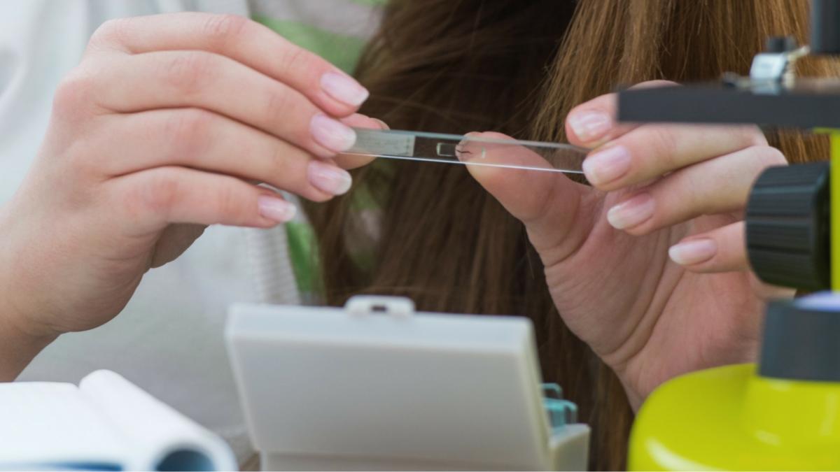
The device allows cellular biologists to view the coverslip directly under the microscope and study cells in great detail. Also, considering that one coverslip can accommodate thousands of tiny elastic stretchers, with each stretcher being able to hold around six cells – that is quite a lot of single cell analysis that you can achieve in a single experiment!
The cell stretcher was made using photolithography-based microfabrication – a broad term for the manufacture of tiny, micro-scaled devices, such as the tiny chips in our mobile phones. “In this process, a chemical solution is poured onto a coverslip, and then UV light is allowed to pass through a ‘stencil’, placed above the coverslip. The light passing through the portion exposed by the stencil, causes the chemicals in the solution to polymerize (forming the rubber-band-like structures), while the rest of the solution is washed away”, explains Mr. Somanna Kollimada, a Senior Research Fellow in the Department of ME at IISc.
The innovative device could be beneficial in the field of cellular biology, and the researchers claim it is easy to manufacture as well. “With this protocol, any biologist can replicate the design with the help of a microfabrication expert”, says Prof. Ananthasuresh.
Today, scientific research is all about establishing inter-disciplinary collaborations, and this study is a prime example in that direction.


